Home / Serviços / Nuclear Medicine

Nuclear Medicine

Functional and metabolic cancer diagnosis (PET-CT and scintigraphy).

Accurate diagnosis for a better clinical decision and more effective follow-up.

Nuclear medicine

What is Nuclear Medicine?

Nuclear Medicine is an advanced medical specialty that uses small amounts of radiopharmaceuticals to diagnose, monitor, and, in some cases, treat different diseases in a precise and minimally invasive way.
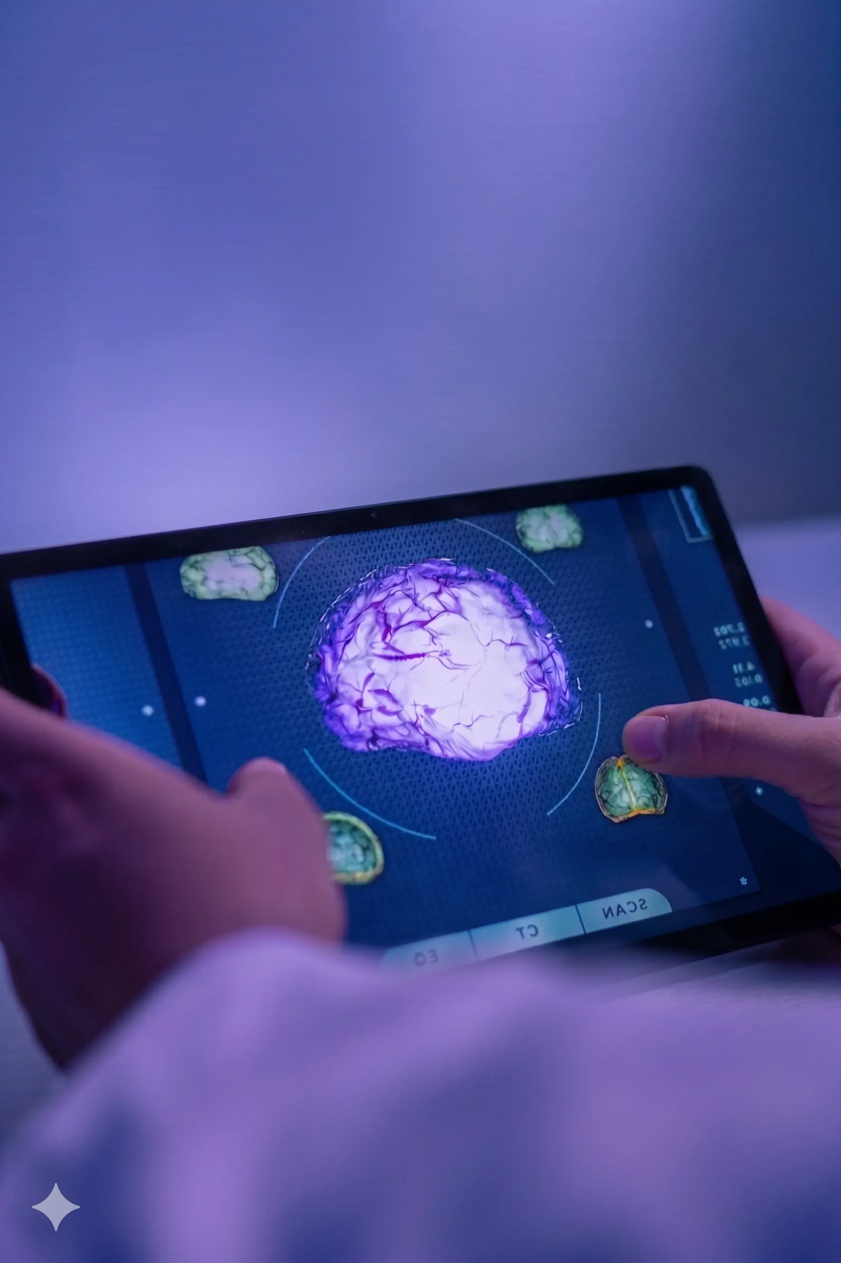
Through functional imaging techniques, such as PET-CT and scintigraphy, Nuclear Medicine allows for the observation of the functioning of organs, tissues, and metabolic processes of the body, facilitating earlier detection and a more precise evaluation of various pathologies, especially in the areas of oncology, neurology, and cardiology.
Pathologies

What pathologies do we study?

Functional imaging and molecular diagnosis in four main areas of clinical application, using PET-CT, SPECT, and scintigraphy.

Benign Pathologies

Detection, staging, follow-up, and evaluation of tumor response.

  • Neurology — Dementia, Parkinson’s, Epilepsy
  • Cardiology — Ischemia and Myocardial Viability
    Endocrinology — Thyroid, Parathyroid
  • Bone Pathology
  • Urology — Isotopic Renogram
  • Gynecology
  • Internal Medicine
  • Infection and Inflammation

Oncological Pathology

Detection, staging, follow-up, and evaluation of tumor response.

  • Breast cancer
  • Prostate cancer
  • Lung cancer
  • Colon cancer
  • Lymphomas
  • Ovarian cancer
  • Cervical cancer
  • Head and neck cancer
  • Melanomas

Can’t find your pathology?

Each case is unique — our medical committee evaluates the clinical indication individually.

Request Evaluation
Diagnosis

Types of tests available

Each Nuclear Medicine test is personalized. Our team adapts the study according to the medical indication and the specific characteristics of each patient.

PET-CT is a non-invasive and painless Nuclear Medicine imaging technique. Unlike conventional X-rays or scans, this test allows for the visualization of not only anatomy, but also the metabolism and functioning of tissues and organs.
It uses positron-emitting isotopes to precisely locate where the injected tracer accumulates, identifying the disease even in its very early stages.

Types of PET

Although the system is a hybrid unit (combining PET with Computed Tomography), there are different applications depending on the radiopharmaceutical used:

  • PET with FDG (Fluorine-18): This is the most common. It uses labeled glucose to detect tumors, infections, and brain abnormalities.
  • Specific radiopharmaceuticals: Advanced tracers are used for greater precision in certain tumors or pathologies, such as:
    • F-Choline and F-PSMA (specific for prostate cancer).
    • Florbetaben or Flutemetamol (for neurology and the study of amyloid plaques in Alzheimer’s).
      Fluorotyrosine, among others.

What is it used for?

Its primary use is to detect where cells have abnormal energy consumption. It is used in:

  • Oncology (85% of cases): For early detection, treatment planning (radiotherapy/chemotherapy), evaluating response to treatment, and detecting recurrence (reappearance of cancer).
  • Neurology: Especially in the diagnosis of Alzheimer’s, epilepsy, and other degenerative diseases.
    Infections and Internal Medicine: Locating hidden infections or issues with prostheses.
  • Cardiology: To study the viability of the heart tissue (myocardium).

How is it performed?

The process is simple and follows these steps:

  1. Injection: The radiopharmaceutical (contrast) is injected into the patient.
  2. Uptake: The patient must wait for a period of time for the cells to metabolize the tracer. Tumor cells, growing faster, consume more glucose (“gasoline”) and accumulate more contrast than healthy ones.
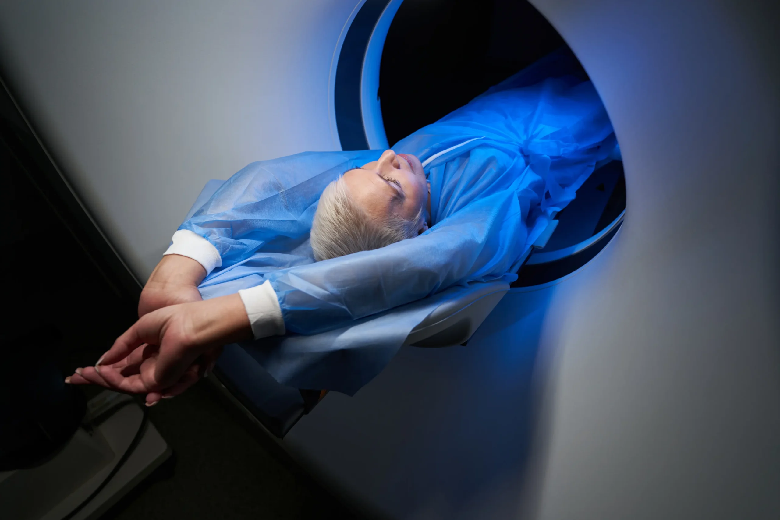
  3. Scanning: The patient lies on the scanner, which detects the radiation emitted from inside the body.
    Image fusion: The system combines the metabolic image (PET) with the anatomical one (CT) to create an exact map of the disease.

Scintigraphy is a Nuclear Medicine imaging technique that provides functional information about organs and tissues. Unlike conventional radiological tests (such as X-ray or CT scan) that show shape or structure (anatomy), scintigraphy shows real-time function and metabolism.

  • It is a safe, non-invasive, and minimally uncomfortable technique for the patient.

Types of Scintigraphy

There are various specialized types depending on the organ or system to be studied:

  • Bone scintigraphy: The most common type, used to detect hidden fractures, infections, inflammatory processes, or bone metastases.
  • Thyroid scintigraphy: Evaluates the function of the thyroid gland and helps diagnose nodules or hyperthyroidism.
  • Lung scintigraphy: Useful for detecting pulmonary embolisms and evaluating blood flow in the lungs.
  • Renal scintigraphy: Studies kidney function and detects possible urinary obstructions.
  • Cardiac scintigraphy: Analyzes blood flow in the heart to diagnose ischemia or heart attacks.
  • Others: Applications in the digestive, lymphatic, and cerebral systems.

What is it used for?

It is mainly used to detect functional abnormalities that are not visible in other tests:

  • Detect hidden inflammatory or infectious diseases.
  • Evaluate the extent of tumor processes (especially in the bones).
  • Study or rule out coronary diseases.
  • Analyze cerebral blood flow or the functioning of organs such as the liver, gallbladder, or kidneys.
  • Confirm or rule out hormonal problems related to the thyroid.

How is it performed?

The procedure consists of four main phases:

  1. Administration of the radiopharmaceutical: A small amount of radioactive material is introduced into the body, usually intravenously.
  2. Distribution time: A specific waiting period (ranging from minutes to hours, or even days depending on the test) is required for the drug to concentrate in the area being studied.
  3. Image acquisition: The patient lies on a table under a gamma camera. This special detector captures the radiation emitted from inside the patient and transforms it into detailed images. It is essential to remain still during this step.
  4. Completion: After the test, the patient should drink plenty of fluids to eliminate the radiopharmaceutical through urine or stool. It is recommended to avoid close contact with children or pregnant women for at least 24 hours.
Equipment

Technology used

Cutting-edge technology for accurate diagnosis and faster turnaround time.

PET-CT Scanners

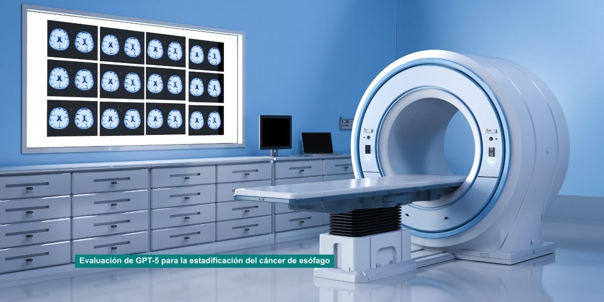
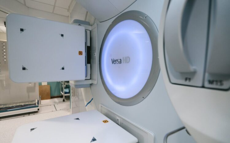
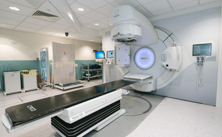
An advanced diagnostic imaging device that combines Positron Emission Tomography (PET) and Computed Tomography (CT) in a single scan.

Gamma Camera/SPECT

A nuclear medicine device that detects radiation and creates detailed images of the inside of the body to diagnose diseases or evaluate organ function

How does it work?

What is the process like?

A structured process to minimise time, guarantee safety and ensure well-coordinated care.

1

Prescription Appointment Request

The PET test always requires a medical prescription. Contact the center chosen to schedule it. Clinical data (usual medication, if diabetic) and insurance data will be requested. You will receive an SMS with the date and time of the exam.

2

Test Preparation

The day before we will confirm your appointment. Come punctually and, if possible, accompanied. You should fast for 6 hours, drink water, avoid intense exercise and consult with our team about the medication you are taking. Report on medical history or diabetes.

3

Conducting the Exam

Bring medical and insurance documentation. Your blood glucose will be monitored at the centre and the radiopharmaceutical will be administered intravenously. Depending on the PET scan, it will be performed immediately or after a few minutes of rest, with an approximate duration of 20 to 35 minutes. Avoid moving and remove metal objects. The test is safe and painless.

4

After the Test

After the exam, you will be able to eat and drink normally. It is recommended to avoid close contact with pregnant women and young children during that day. Continue to drink fluids to promote the elimination of the radiopharmaceutical.

5

Results

The results are not immediate, as the images must be processed and analysed by specialists in Nuclear Medicine. The centre will inform you about the different means available to access your study.

1

Prescription Appointment Request

The PET test always requires a medical prescription. Contact the center chosen to schedule it. Clinical data (usual medication, if diabetic) and insurance data will be requested. You will receive an SMS with the date and time of the exam.

2

Test Preparation

The day before we will confirm your appointment. Come punctually and, if possible, accompanied. The centre’s staff will give you the necessary instructions on whether you should fast or withdraw any medication, depending on the type of scintigraphy. Report on medical history, pregnancy, allergies, infections or cancer treatments.

3

Conducting the Exam

Bring medical and insurance documentation. The radiopharmaceutical will be administered intravenously at the centre, and the scintigraphy or SPECT will be performed immediately or after waiting a certain time. Avoid moving and remove metal objects. The test is safe and painless.

4

After the Test

After the exam, you will be able to eat and drink normally. It is recommended to avoid close contact with pregnant women and young children during that day. Continue to drink fluids to promote the elimination of the radiopharmaceutical.

5

Results

The results are not immediate, as the images must be processed and analysed by specialists in Nuclear Medicine. The centre will inform you about the different means available to access your study.

Comprehensive support throughout the process

In addition to medical treatment, our team is available to answer questions, manage symptoms and accompany you at every stage. Radiotherapy is a path that we travel together.

Our differential value

Why Choose Us?

We combine cutting-edge technology with a people-centred care model.

  • Advanced
    technologyState-of-the-art equipment with millimeter precision control in each treatment session.

  • Agile
    planningFirst preferential visit and initiation of treatment without delay

  • Personalized
    attentionEach treatment plan is designed specifically for the patient, according to the type of tumor, its location and clinical situation.

  • Multidisciplinary
    teamsOncologists, radiophysicists, technicians and specialized nurses work in coordination in each clinical case.

  • Reduced displacement
    Service available in nearby reference hospitals, avoiding long transfers at a delicate time.

  • Hospital
    integrationWe work integrated into the hospital circuit, guaranteeing fluid coordination with the rest of the specialists.

  • Quality and clinical
    safetyWe follow up-to-date international protocols with continuous verification of safety at every stage of treatment.

Where we are

Alliance Medical Centers with Nuclear Medicine Unit

Alliance Medical – Córdoba

Hospital San Juan de Dios, Av. del Brillante, 106, 14012 Córdoba

Mon–Fri 8:00 a.m. to 4:00 p.m.

+34 957 85 86 69

Alliance Medical – Granada

Pol. Ind. Juncaril – Parcela 102, 18210 Peligros, Granada

Mon–Fri 8:00 a.m. to 4:00 p.m.

+34 958 40 18 13

Alliance Medical – Jeréz de la Frontera

Calle Newton 13, 11407 Jerez de la Frontera, Cádiz

Tuesday 8:00 a.m. to 4:00 p.m. — Mon-Fri 8:00 a.m. to 4:00 p.m.

+34 856 56 59 84

Alliance Medical – La Línea de la Concepción

Carretera Av. del Higuerón, 109, Pol. Zabal Bajo 11300 La Línea de la Concepción, Cádiz

Mon–Fri 8:00 a.m. to 4:00 p.m.

+34 956 90 06 16

Alliance Medical – Sevilla

Hospital de la Cruz Roja, Ronda de Capuchinos, nº 11 · 41009 Sevilla

Mon–Fri 8:00 a.m. to 4:00 p.m.

+34 954 43 25 37

Alliance Medical – Málaga

Avenida Andalucía, Nº 27 (bajo) 29006 Málaga

Mon–Fri 8:00 a.m. to 4:00 p.m.

+34 952 36 38 91

Who are we addressing?

Select your profile and we'll guide you through the process

We have two different forms to speed up the process: one for patients and another for doctors who wish to request an appointment directly.

I'M A PATIENT

Request an appointment

We help you find the most suitable service and center for your case.

Request an appointment online No account required · Instant confirmation
I'M A PATIENT

Refer a patient

Access the referral form and submit the relevant clinical documentation.

Refer a patient online No account required · Instant confirmation
Medical area

Much more than medicine: content for your everyday life

Go to News and Content

Evaluación de GPT-5 para la estadificación del cáncer de esófago

Evaluar si un modelo de lenguaje grande multimodal (GPT-5) puede interpretar imágenes de PET con FDG y ayudar a estadificar el cáncer de esófago, comparando…

Read more

Alliance Medical adquiere las actividades ibéricas de Mercurius Health Group

Alliance Medical, el principal proveedor independiente de servicios de diagnóstico por imagen en Europa, anuncia que ha concluido un acuerdo para adquirir las actividades ibéricas…

Read more

(No) te lo tomes como un hombre

Expresiones como «tómatelo como un hombre» o «sé un hombre» están (todavía) muy presentes en nuestra sociedad. Quizás sin darnos cuenta, al utilizar estas expresiones…

Read more

El Hospital Viamed Tarragona tendrá una Unidad integral de Oncología y Radioterapia avanzada para posicionar la  provincia como referente en el abordaje del cáncer 

El nuevo Hospital Viamed Tarragona se prepara para abrir sus puertas con una firme apuesta por la excelencia asistencial en el ámbito oncológico. Actualmente, se…

Read more

Cáncer de mama

Hay mucho que decir sobre el cáncer de mama. Al ser uno de los cánceres más comunes del mundo, existen libros enteros sobre el tema,…

Read more

Importancia de los datos de radioterapia

La gestión eficaz de los datos médicos es especialmente crítica en radioterapia, ya que los datos son necesarios para cada tratamiento. Sin acceso en línea…

Read more

¿Cómo funciona la radioterapia?

Siendo el objetivo de la radioterapia matar las células cancerosas, permítame que le lleve en un recorrido microscópico por el interior de una sola célula…

Read more

Inteligencia artificial en oncología

La Inteligencia Artificial, también conocida como IA, es el desarrollo de aplicaciones informáticas para realizar tareas que normalmente requieren inteligencia humana, como la comprensión del…

Read more

La ciberseguridad se ha convertido en un problema crítico de seguridad sanitaria tras el creciente número de incidentes de gran repercusión.

La ciberseguridad de los dispositivos médicos es una de las principales preocupaciones actuales. En el entorno médico actual, casi todos los dispositivos médicos incluyen algún…

Read more
Asociaciones

John R. Adler: “Mi objetivo es tratar cada año a más de 4 millones de pacientes que ahora mismo no tienen una opción de tratamiento de radiocirugía”

Madrid, 22 de marzo de 2023. John R. Adler, figura mundial de la radiocirugía, ha visitado España para conocer las instalaciones del Instituto de Radiocirugía Avanzada (IRCA), ubicadas…

Read more

Frequently Asked Questions

Once there is the slightest suspicion of a possible tumour lesion, it is the most effective technique and the one that will provide more information compared to other diagnostic tests, providing the specialist with a better initial assessment of the disease and a subsequent more effective planning.

The amount of radiation emitted and received by the patient is extraordinarily low, similar to that received during an abdominopelvic scan. The injected amount of radiopharmaceutical is so small that there is no risk of any type of reaction. The only contraindications are: pregnant women and people with extreme obesity who may have difficulty accessing the scanner. If in doubt, always consult the specialists at the PET Center.

Yes. This test must always be prescribed by a physician, and it will normally be to them that you must submit the results. Your doctor must have reasons to believe that this technique will be useful for you, in accordance with the existing indications for its use.

We currently have service agreements with the leading medical companies in the national and European market. Depending on the type of policy you hold, this service may or may not be covered, so you should check with your health insurance company beforehand.

Once your doctor has prescribed the test, you can request your appointment by contacting the center by phone or email. Our team will let you know the date and time, answer any questions, and request the necessary information from you. You will also receive a confirmation of your appointment by SMS.

The day before your exam, our team will contact you to confirm the appointment time and answer any additional questions. It is recommended that you avoid physical exertion before coming to the center and, if possible, come accompanied by an adult. The PET/CT scan is a simple, safe test that is well tolerated by patients.

Fast for 6 hours before the test, avoiding solids and sugary liquids. Stay hydrated from the day before your appointment. You may take a tranquilizer if needed, and if you have any doubts about medication, please contact us. Do not engage in intense physical exercise in the days leading up to the test.

Yes. Our staff will give you a form on which you must indicate whether you have had any type of tumor; whether you have undergone surgery; whether you are pregnant or could be, or are breastfeeding; whether you have any allergy to medication or contrast; whether you have or have recently had an infection; whether you have recently received chemotherapy or radiotherapy; whether your weight is over 100 kg; and if you are diabetic, you must have controlled blood glucose levels (if the diabetes is insulin-dependent, you should not receive insulin for 4 to 6 hours before the test).

Company documentation: company authorization, health insurance card, and doctor’s prescription. Medical documentation: reports and images (CT, MRI…) that you have not previously submitted, and the report from the doctor who ordered the test. ID card and health insurance card.

It is very important that you arrive on time for your appointment and are very punctual, as the isotopes used for this test have a short lifespan and disappear quickly. Excessive delays could result in the test having to be postponed to another day.

Once at the center, your height and weight will be measured and your blood glucose level will be checked. A venous line will then be inserted for the administration of the radiopharmaceutical. The injection is completely painless and harmless, with no known side effects or risk of allergies. After the injection, you will need to lie down comfortably in a room for 45 minutes while the radiopharmaceutical is properly distributed throughout the body.

After the 45-minute period for the radiopharmaceutical to take effect, you will proceed to the PET scanner. The scan lasts between 20 and 35 minutes depending on the type of study. You must remain still once the study has begun and remove any metal objects. The test carries no risk or side effects.

Once the study is complete, you can eat and drink normally. It is advisable not to get close to pregnant women or young children on the day of the test, as they are more sensitive to radiation. By the next day, the radiopharmaceutical will have been completely eliminated and you can resume your normal life. Continue drinking plenty of fluids throughout the day.

The results are not available immediately after the test. The information obtained must be processed by powerful computers in order to produce the images that will be evaluated by Nuclear Medicine specialists. The center will inform you of the different ways to access your study.

Need more information?

Contact our team or request your appointment easily.

This site is registered on wpml.org as a development site. Switch to a production site key to remove this banner.